
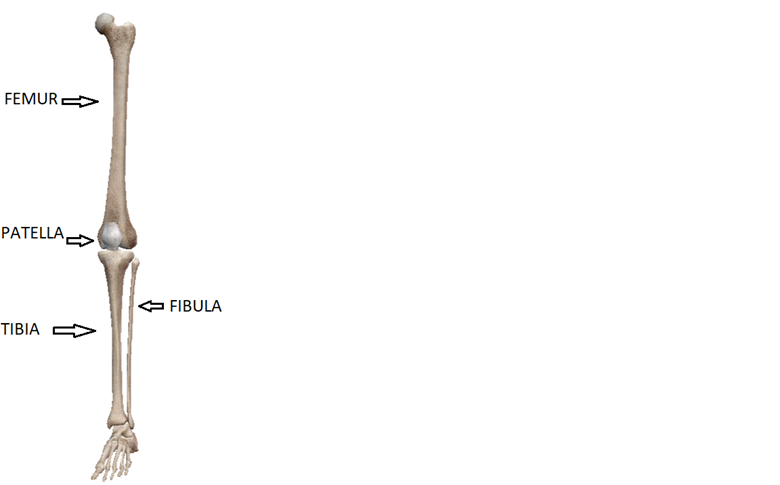
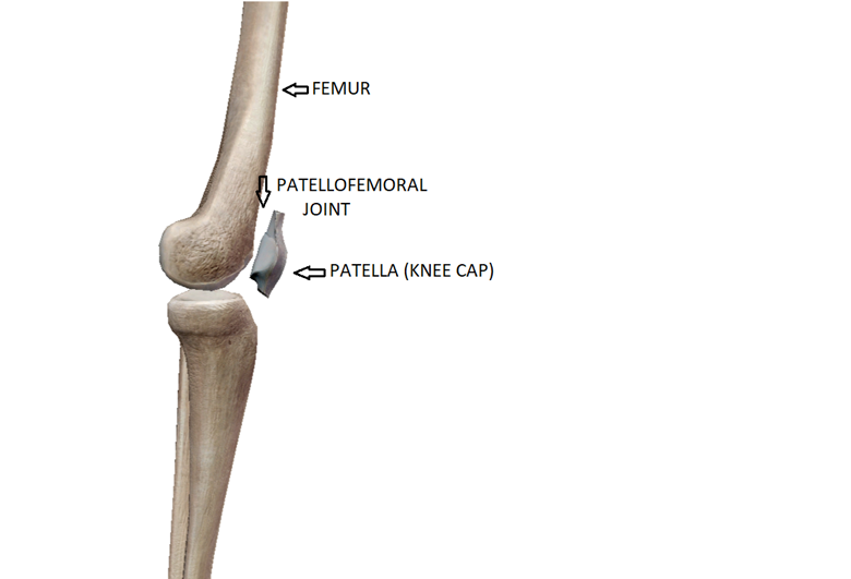
The knee is a hinge joint between the hip and ankle in the leg. The knee is a synovial joint consisting of 4 bones including the femur, tibia, patella, and fibula.
Joint
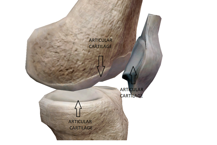
A joint is where two or more bones meet and move (articulate) on one another. The main articulations of the knee are between the femur and tibia (tibiofemoral), and the femur and patella (patellofemoral). The articulating surfaces of bones are covered with a layer of hyaline cartilage also called articular cartilage. This cartilage provides a smooth almost frictionless surface for the bones to move on one another.
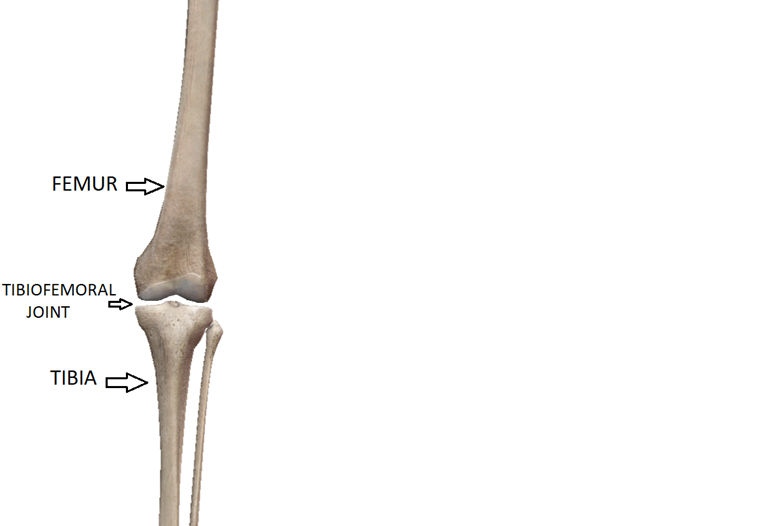
Tibiofemoral Joint
The tibiofemoral joint of the knee is formed by the distal end of the femur and proximal end of the tibial.
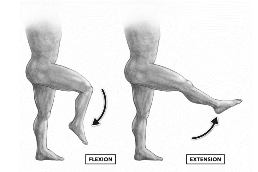
The main motions of this joint are flexion and extension, but also include medial and lateral rotation. The average range of motion for the knee is 0-135 degrees.
Patellofemoral Joint
The patellofemoral joint of the knee is formed by the convex patella and the concave groove between the femoral condyles. The patella glides in this groove of the femur during knee movement and functions to not only protect the inside of the joint, but also increases the strength of the extensor mechanism by 30-50%.3
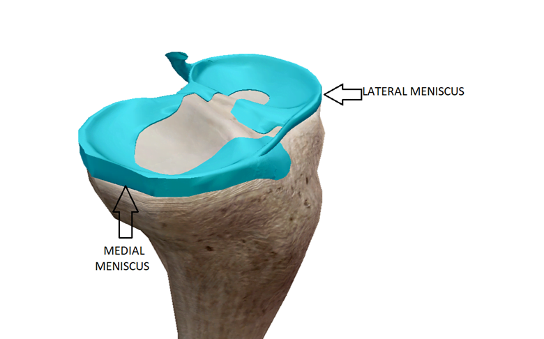
Meniscus (Menisci)
The knee has two c-shaped rubbery fibrocartilage structures that wrap around and attach to the periphery of the tibia. The menisci are thicker at the periphery and thinner at the inside edges. The inside edges of the menisci do not attach to the tibia which allows for some movement. The meniscus deepen the articular surface of the tibia to increase joint congruency and stability with the femur, as well as provide shock absorption and contribute to lubrication and nutrition.
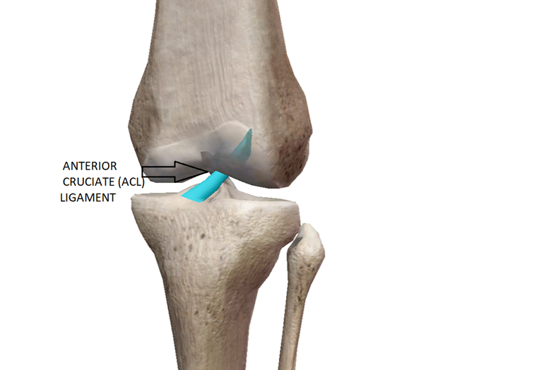
Ligaments
Ligaments are fibrous connective tissue that attach bone to bone. They promote proper joint mechanics, as well as prevent excessive joint movement. Below are the 4 major ligaments of the knee.
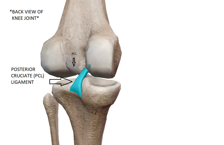
Anterior Cruciate Ligament (ACL)
The ACL attaches from the femur to the tibia and runs diagonally inside the knee joint making an “X” with the posterior cruciate ligament. It prevents the tibia from moving too far forward on the femur and provides rotational stability.
Posterior Cruciate Ligament (PCL)
The PCL attaches from the femur to the tibia and runs diagonally inside the knee joint in the opposite direction as the ACL to make an “X”. It prevents the tibia from moving too far behind the femur and provides rotational stability.
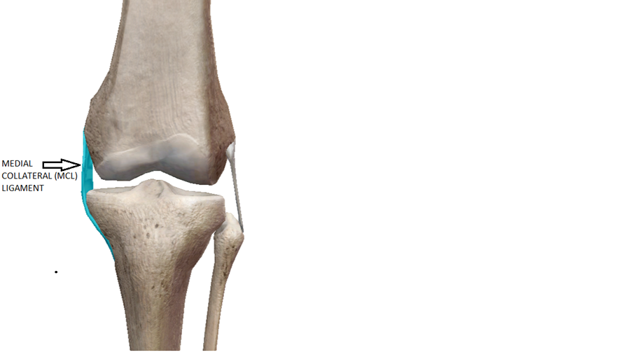
Medial Collateral Ligament (MCL)
The MCL attaches from the femur to the tibia on the inside of the knee. It prevents excessive valgus, or “knock knee” movement in the knee.
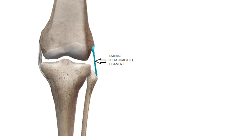
Lateral Collateral Ligament (LCL)
The LCL attaches from the femur to the fibula on the outside of the knee. It prevents excessive varus, or “bowed knee” movement in the knee.
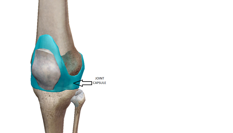
Joint Capsule
The joint capsule is a fluid filled layer of fibrous connective tissue that surrounds the entire joint and encapsulates it. The joint capsule provides joint stability, proprioception, and prevents excessive movement in all directions. It is also essential for joint nutrition and lubrication as the inside layer of the capsule has a synovial membrane that secretes synovial fluid.
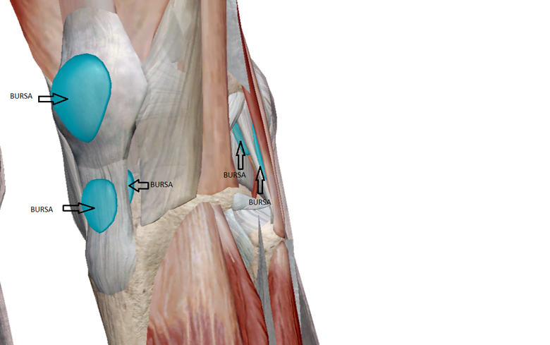
Bursa (Bursae)
Bursae are small fluid-filled sacs that rest in between moving parts near joints and reduce friction thus preventing tissue wearing. Below are a major bursae of the knee:
Suprapatellar bursa
Infrapatellar bursa
Pes Anserine bursa
Medial collateral ligament bursa
Iliotibial bursa
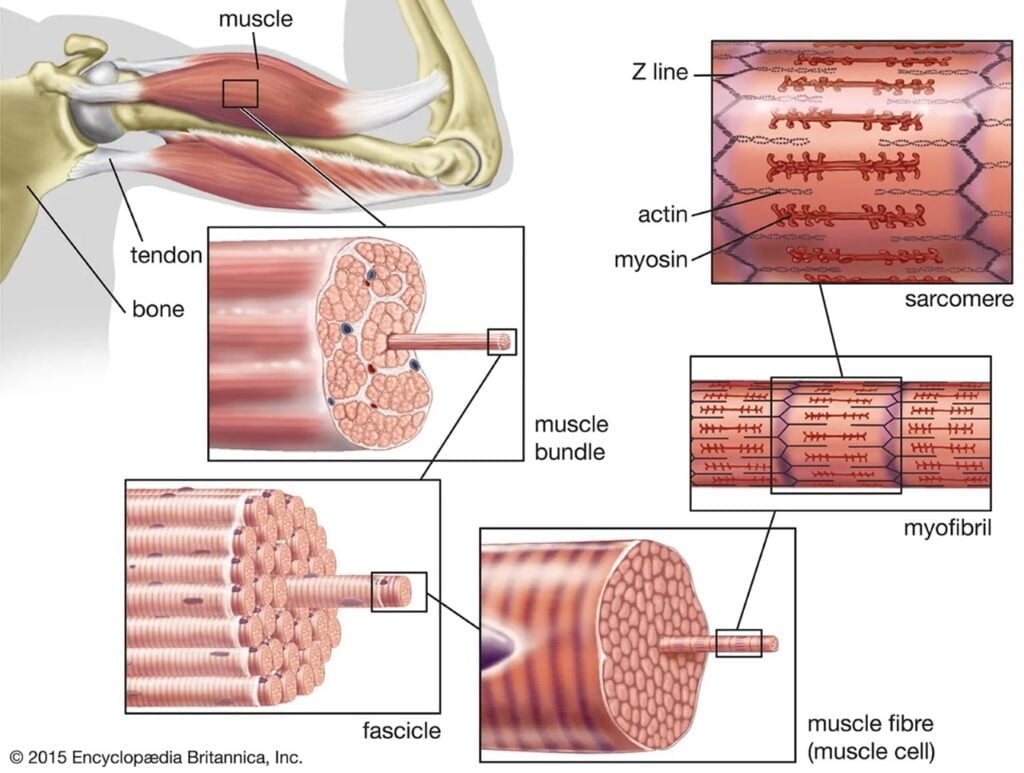
Muscle(s) and Tendon(s)
Skeletal muscle is made of bundles of contractile fibers that produce movement. The muscle attaches to bone via a tendon which is made of fibrous connective tissue. All skeletal muscles have a proximal and distal tendon, and when the muscle contracts/shortens it pulls the attached bones closer together. Muscles not only produce bodily movement, but also control movement and stabilize joint(s) and posture(s). Below are important muscles and tendons of the knee:
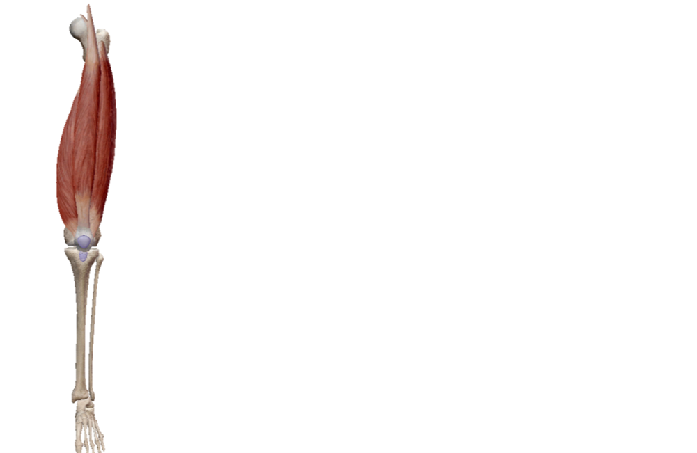
Quadriceps
The quadriceps are a large group of four muscles (rectus femoris, vastus lateralis, vastus medius, vastus intermedius) on the front of the thigh. The muscle action of the quadriceps is flexion of the hip, extension of the knee, and provides patellar stability.
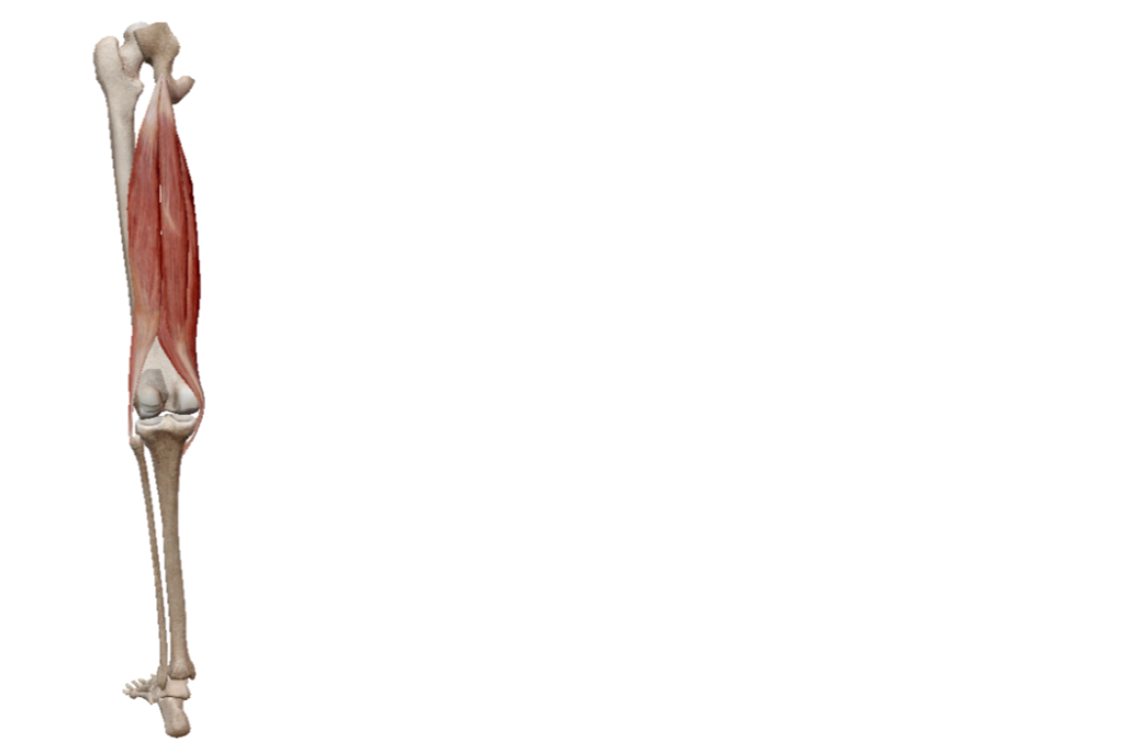
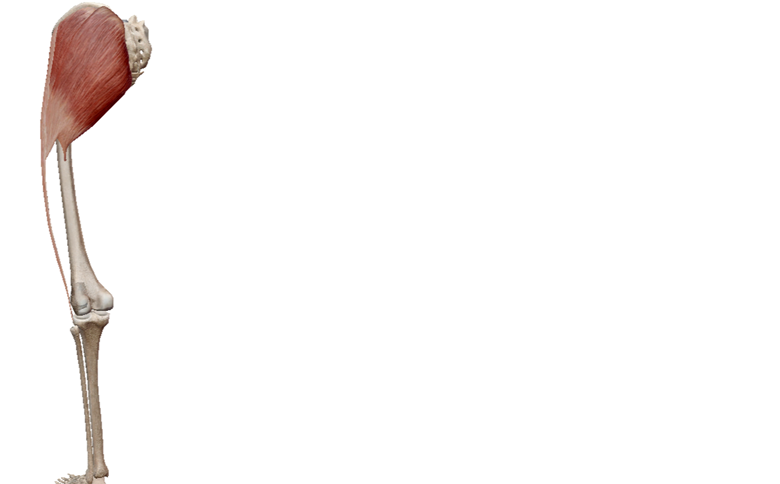
Hamstrings
The hamstrings are a large group of three muscles (biceps femoris, semimembranosus, semitendinosus) on the back of the thigh. The muscle action of the hamstrings is extension of the hip and flexion of the knee.
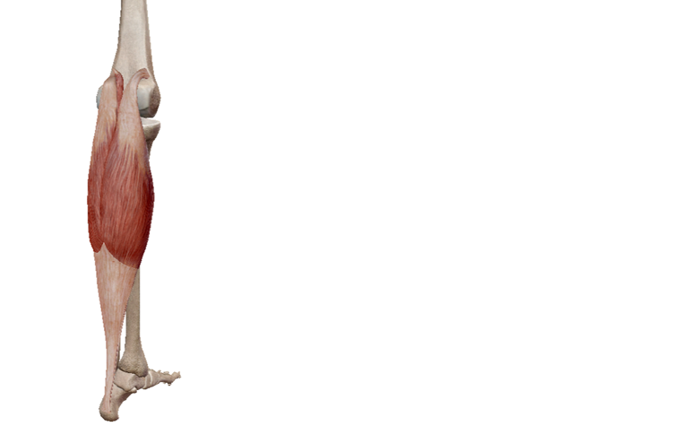
Gastrocnemius/Plantaris
The gastrocnemius/plantaris are muscles of the calf that cross the knee and assist the hamstrings with knee flexion.
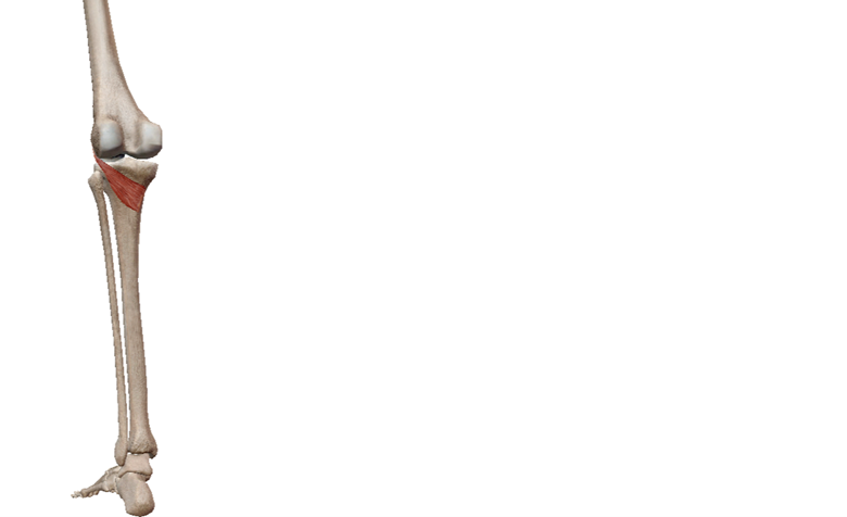
Popliteus
The popliteus is a small muscle found deep in the back side of the knee (popliteal fossa). The muscle action of the popliteus is to unlock the knee as it bends and assist with joint stability.
Gluteal Muscles/Tensor Fasciae Latae-Iliotibial Band
These muscles wrap around the lateral and back side of the hip, but have a profound influence on the knee joint. The gluteal muscle group (gluteus maximus, gluteus medius, gluteus minimus) stabilize the knee by controlling femoral position and movement. The Tensor fasciae latae and the iliotibial band also provide lateral stability to the knee.
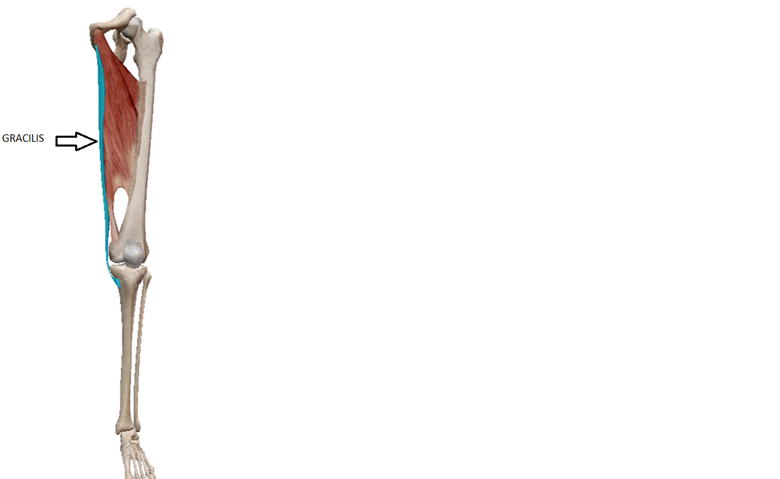
Adductor Muscles
The adductors (adductor magnus, adductor longus, adductor brevis, and gracilis) are a group of muscles on the inner thigh. The gracilis is the only adductor that crosses the knee joint and its muscle action adduction of the hip, flexion of the knee, and medial rotation of the tibia.


0 Comments